


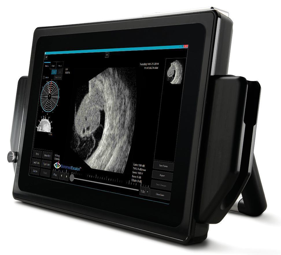


VuPad's superior UBM and B-scan image quality with Enhanced Focus Rendering and ultra high resolution touch screen allows you to capture both crisp still images and record video that can be carefully reviewed frame-by-frame.
Exceptional User Experience
Concentrate on your patients, not on the controls. With an intuitive graphic interface, VuPad™ makes ultrasound simple and straightforward. The multi-touch screen puts important functions easily at your fingertips. You can also take advantage of innovative, smartphone-inspired features like pinch zoom. VuPad™ also includes time saving pre-set scan settings to automatically optimize image quality depending upon area of viewing interest.
Portable. Flexible. Adaptable.
In ophthalmic practices, there’s no such thing as an “ordinary day”. The compact, ergonomic VuPad is designed to adapt. You choose the modalities that you want – UBM, B-scan, A-scan and/or pachymetry. Dual-Band WIFI, Ethernet, USB, and Bluetooth allow you connect to other devices or your network. Large high resolution touchscreen with HDMI output. There’s also plenty of room onboard to store exams with a customizable high-performance SSD drive and the most flexible interconnectivity capabilities, including DICOM and several export options
Advanced Angle Analysis
Allows accurate quantification and tracking of angle properties, including differences during mydriatic and miotic conditions.
Intuitive, Efficient Workflow
Quickly perform and review ultrasound exams with easy to use touch interface, preset scan modes to effortlessly optimize image quality for area of interest, frame-by-frame review of up to 12 video clips, use of touch pinch zoom, and more.
Eye Tracking Alignment
Provides real-time feedback to ensure proper alignment of UBM scans for sulcus-to-sulcus measurements.
Connected - Integrated
Easily connect VuPad to your network, wireless keyboard, external monitor, EHR, and/or PACS